Bone strength tends to decline gradually, often without clear warning signs, until a fracture occurs. In clinical settings, many cases emerge after minor falls, revealing underlying bone loss that developed over the years. Osteoporosis places pressure on healthcare systems due to delayed diagnosis and limited early screening uptake. Daily habits influence bone remodeling, yet adherence remains inconsistent. Targeted lifestyle adjustments, combined with awareness of clinical limitations, can reduce fracture risk and support long-term skeletal stability in both aging and at-risk populations.
6 Practical Strategies to Support Bone Density and Reduce Osteoporosis Risk
Maintain Adequate Calcium Intake Through Diet
Calcium intake often looks fine on paper, then a food diary tells another story. Clinic diet histories commonly show long stretches of low-calcium meals, especially among older adults eating the same few staples. Milk, yogurt, cheese, canned salmon with bones, tofu set with calcium, leafy greens, and fortified cereals can fill the gap, but absorption is not uniform.
Acid-suppressing medicines, bowel disease, and bariatric surgery can reduce uptake, so dietitians may spread calcium-rich foods across the day instead of relying on one large serving. Supplements can help, yet dosing needs care in people with kidney stones, chronic kidney disease, or high blood calcium.
After hospital discharge, written instructions are easy to miss. Practical plans work better: simple grocery lists, measured portions, and follow-up checks during routine visits. Lab results and medication lists should be reviewed together, since some therapies interfere with calcium balance long-term.
Ensure Sufficient Vitamin D Levels for Absorption
Vitamin D shortages show up often on routine bloodwork, particularly during long winters or in people spending most days indoors. Low levels blunt calcium uptake in the gut, so the body pulls calcium from bone to keep blood levels steady. That silent tradeoff can surface later as a wrist fracture after a small slip, or as repeated stress injuries.

Clinics usually confirm status with a 25-hydroxyvitamin D test, yet borderline results sometimes get sidelined when visits focus on diabetes, blood pressure, or pain. Clear deficiency is treated more decisively, often with a short course of higher-dose tablets followed by a maintenance dose.
Sun exposure can help, but age, darker skin, sunscreen use, and limited mobility reduce production. Supplements offer predictable dosing, but kidney disease, sarcoidosis, and certain medications can change targets. Follow-up labs prevent overshooting and keep plans grounded. Food sources remain limited, so diet alone rarely corrects low values.
Engage in Weight-Bearing Physical Activity
Mechanical stress stimulates bone formation, making weight-bearing activity a key factor in maintaining density. Physical therapy departments often incorporate these exercises into rehabilitation plans following fractures. Walking, stair climbing, and resistance training form the core of most programs.
A common barrier involves joint pain or mobility limitations, particularly in patients with arthritis. In such cases, therapists adapt routines to reduce strain while preserving bone-loading benefits. For example, short intervals of standing exercises may replace longer walking sessions.
Adherence remains a challenge outside supervised settings. Many individuals discontinue activity after initial improvement, reducing long-term benefit. Community-based programs sometimes help bridge this gap, though access varies. Tracking progress through simple metrics, such as step counts or resistance levels, can provide structure and maintain engagement over time.
Limit Factors That Accelerate Bone Loss
Bone loss can speed up quietly when certain exposures stack up. Chronic corticosteroid therapy is a common trigger; rheumatology and pulmonary clinics often see patients on repeated bursts for flares, and bone protection can lag behind refill cycles.
Smoking further suppresses bone-building cells, and heavy caffeine use can nudge calcium balance in the wrong direction, especially when meals are irregular. A practical safeguard is a routine medication review. Pharmacists can spot long-term steroids, high-dose thyroid replacement, some seizure medicines, or acid suppressants, then alert prescribers to screening needs or alternatives.
The main limitation is fragmented care, with prescriptions coming from multiple offices. Shared records and a clear follow-up plan reduce missed opportunities. During annual visits, pairing lifestyle notes with lab checks and DXA scheduling keeps prevention from becoming an afterthought.
Monitor Bone Density Through Screening
Bone density testing, typically performed using dual-energy X-ray absorptiometry, offers a measurable way to assess fracture risk. Screening guidelines vary, which leads to inconsistent testing across populations. Many individuals receive scans only after a fracture, limiting opportunities for early prevention.
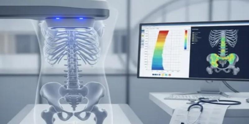
In clinical settings, scheduling delays and insurance coverage issues often affect access. Some patients decline testing due to lack of symptoms, despite underlying risk factors such as family history or long-term medication use. Education during routine visits can help address these gaps, though time constraints remain a barrier.
Results from bone density scans guide treatment decisions, including medication initiation. However, follow-up testing is sometimes overlooked, making it difficult to track progression. Integrating reminders into health systems improves continuity, allowing clinicians to adjust care plans based on measurable changes over time.
Incorporate Protein and Balanced Nutrition
Protein gives bones their scaffolding, and intake often slips as meals get smaller with age. On hospital wards, diet screens often show low protein alongside poor grip strength and unsteady walking, a mix that raises fall and fracture risk.
Care plans pair protein foods with calcium, vitamin D, and produce to cover minerals, energy, and fiber. Recovery periods can derail eating. Sore gums, ill-fitting dentures, nausea from new medications, or fatigue after infection can turn full plates into leftovers.
In those moments, clinicians may add oral nutrition supplements, then reassess to avoid replacing meals long term. Dietitians coordinate with nursing staff and family caregivers, using snack schedules, texture changes, and protein at breakfast to keep intake steady at home. Simple logs of intake help spot dips before weight loss appears.
Conclusion
Bone health reflects long-term patterns shaped by diet, activity, and clinical care. Small gaps in daily habits often accumulate, leading to measurable loss over time. Clinical interventions provide structure, yet sustained improvement depends on consistent follow-through outside healthcare settings. Screening, nutrition, and physical activity each address different aspects of bone stability. When applied together, these approaches reduce fracture risk and support functional independence. Ongoing monitoring ensures adjustments remain aligned with changing health needs and individual risk factors.